Case of the Month - October 2024
B - Malignancy
SVC syndrome is most often caused by malignancy with compression of the SVC related to external compression, lymphadenopathy, or direct venous invasion. However, benign etiologies such as fibrosing mediastinitis, existing central venous catheters, in-stent re-stenosis, and implanted cardiac devices are estimated to account for up to 40% of SVC syndrome cases. Treatment may include systemic steroids, radiotherapy, and angioplasty with or without stent placement. Surgical bypass is another option but rarely performed due to the higher morbidity and mortality particularly since many patients with malignancy SVC syndrome are poor surgical candidates.
Data on stenting for SVC syndrome is limited and heterogeneous. Generally, there is clinical improvement in >90% of cases with reported one year patency rates ranging from 60-100%, though most report 80-90%. Decreased patency/recurrent obstruction is more common with malignant obstruction, longer obstructive lesions, and those associated with a persistent endovenous device such as a stent, device lead, or catheter.
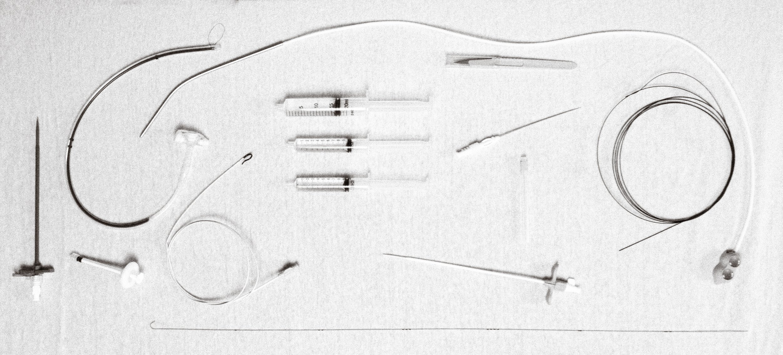
To treat the recurrent obstruction, right common femoral access was obtained. For chronic obstructions, a tri-axial system is often required to provide enough support to cross the obstructing lesion. A long 8 Fr sheath was used for access through which a 7 Fr hockey stick and 5 Fr Kumpe catheter were used to create a tri-axial system for crossing the obstruction. For the patent caudal portion of the stent, a J-tipped Rosen wire was used to not cannulate the stent interstices. An angeled glidewire was then used in combination with the tri-axial system to cross the obstructing lesion. Initial venography was then performed.
Which of the following findings is seen in the initial venography below?
A. Patent existing stent
B. Focal thrombus at the existing port catheter tip
C. Right atrial opacification
D. Stent obstruction with filling of tortuous venous collaterals

